


Ready to care for your smile with confidence.
For more information or to schedule a consultation with a dentist at Global Dental Complex, please contact us.
📞 Call / WhatsApp: (+66) 065-669-9191
💬 LINE: @globaldentalcpx
🌐 Website: globaldentalcomplex.com/th
Book your consultation today and start planning the treatment that is right for you.
Advanced Dental CT Scan (CBCT) & Digital X-ray in Bangkok
What is a CBCT Scan (3D Dental X-ray)?
Visual your problem more clearly. Help your doctor to provide more accurate treatment.
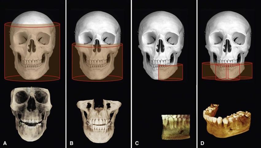
CBCT scans capturing data using a cone-shaped X-ray beam. CBCT is used in dental profession. These data are used to reconstruct a three-dimensional (3D) image of the following regions of the patient’s anatomy: dental (teeth); oral and maxillofacial region (mouth, jaw, and neck); and ears, nose, and throat (“ENT”). Also, the CBCT data can create 3D volumetric rendering.
Dental CT (CBCT) vs. Medical CT Scan
- CBCT is better than the conventional CT used in medical field because it is less expensive and has lower doses of absorption.
- The CBCT systems used by dental professionals rotate around the patient, capturing data using a cone-shaped X-ray beam.
What is the benefits of CBCT?
- dental CBCT, provides a fast, non-invasive way of answering several clinical questions.
- Dental CBCT images provide three-dimensional (3-D) information, rather than the two-dimensional (2-D) information provided by a conventional X-ray image.
- 2D imaging had some weakness such as image enlargement, elongated or shortened object, image distortion, superimposed structures. The real anatomy could be evaluated and analyzed better from three-dimensional (3D) image.
What is the advantage of CBCT over 2D image?
- reliable diagnosis tool for the detection and quantification of bone resorption in periodontal diseases.
- Images without magnification
- Better visualization of the defective
- No distortion and overlapping of the images from CBCT.
Due to present better detail, CBCT is used in many ways.
- Locate the impacted or retained teeth.
- Locate impacted tooth in order to place bracket for orthodontic treatment.
- Treatment planning for dental implant
- Surgical simulation for orthognathic surgery case (Surgery)
- Find the amount of root canals.
- Evaluate the bone height and thickness.
Digital X- Ray
A digital x-ray allows the dentist to take an image of the tooth or teeth and put it into an imaging program immediately. Within this imaging program, there are several tools that will allow the dentist to take a close look at the teeth and surrounding structures with amazing accuracy. As a benefit to the patient, the digital x-ray also provides nearly 80% less radiation than a standard x-ray. This is because the digital version of the x-ray is much more sensitive to this radiation and has been specifically designed with the patient in mind.
Procedure of digital dental X-ray
The actual procedure for having a digital dental x-ray is very similar to a traditional x-ray. our highly trained dental team will carefully insert a sensor into your mouth, and which will capture the image of your teeth or jaws. This digital sensor sends the information directly to a computer so that the images taken can be instantly viewed on a screen in the treatment room. Alternatively, images may be taken using a specialized scanner, or we can use a combination of a sensor and a scanner to produce a digital image. The exact method used is dependent on the type of x-ray required.
Key Benefits of Digital Dental X-rays
- Once the digital image is on-screen, we can adjust it if necessary, enlarging or magnifying any areas which require close inspection. This makes it much more straightforward to identify any small cavities or other areas of concern.
- Diagnose any problems more efficiently and more quickly,
- Be available for viewing immediately since it does not have to be first developed like the old film x-rays.
- It is also very straightforward for us to show you these images on-screen and to explain any problems and how best to treat them.
- If a second opinion is required, these digital images can be emailed to a dental specialist, a process which is much faster and more efficient than having to mail conventional x-ray films.
- Available for patient education
Types of Digital Dental X-rays We Offer
Digital dental x-rays can be taken inside your mouth (intraoral), or they may be taken outside your mouth (extraoral).
Intraoral x-rays include:
- Bite-wing X-Rays: You must bite down on a sensor for an x-ray can be taken in a specific part of the mouth. Bite-wing x-rays are frequently used for detecting decay in between teeth, to check the condition of bone around teeth and to assess the fit and integrity of dental restorations including crowns and fillings.
- Periapical X-Rays: A periapical x-ray shows the entire tooth from its crown to the tip of the tooth root, as well as the bone surrounding the tooth. Periapical x-rays are useful in detecting periapical lesion and for assessing bone loss around the tooth which can occur if you have advanced gum disease.
Extraoral x-rays include:
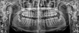
- Panoramic X-Rays: A panoramic x-ray is taken by a machine that rotates around your head, providing a single detailed image of all your teeth in your upper and lower arch. Panoramic x-rays are particularly useful for assessing impacted wisdom teeth and other jaw problems, and for planning dental treatments including implants.
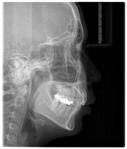
- Lateral Cephalometric x-rays: is particularly useful for orthodontic treatment planning.
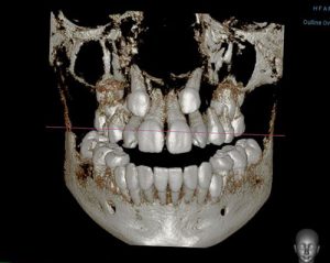
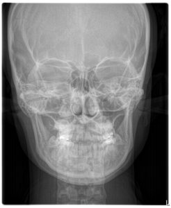
- Postero-Anterior Cephalometric x-rays is particularly useful for identify facial asymmetry in orthognathic surgery case.
Dental CT Scan & X-Ray: Accurate Dental Imaging for Better Diagnosis and Treatment Planning
Many dental problems cannot be seen with the eyes alone. Cavities between teeth, infections around tooth roots, impacted wisdom teeth, bone loss, jaw problems, and hidden dental structures often require imaging for accurate diagnosis. Dental CT scan & x-ray services help dentists see what is happening beneath the surface, allowing for safer, more precise, and more personalized treatment planning.
Dental imaging may include small intraoral X-rays, panoramic X-rays, cephalometric X-rays, and 3D dental CT scans such as CBCT, or cone beam computed tomography. Each type of imaging has a different purpose. A simple X-ray may be enough for a cavity or root infection, while a 3D dental CT scan may be recommended for implants, wisdom teeth, jaw surgery, complex root canal cases, or bone evaluation.
A Dental CT scan & x-ray should be used only when clinically necessary. The dentist will choose the most appropriate image based on your symptoms, oral condition, treatment plan, and the level of detail needed.
What Is a Dental X-Ray?
A dental X-ray is a diagnostic image that helps dentists see teeth, roots, bone, and surrounding structures that are not visible during a normal examination. Dental radiographs can help detect cavities, bone loss, dental infections, tooth development problems, and other hidden conditions.
Dental X-rays are commonly used during check-ups, emergency visits, root canal diagnosis, periodontal evaluation, orthodontic planning, and before certain dental procedures. They provide important information that supports diagnosis and treatment decisions.
A dental X-ray is usually quick and non-invasive. Digital X-ray systems allow images to appear on a screen soon after capture, making it easier for the dentist to explain findings to the patient.
What Is a Dental CT Scan?
A dental CT scan, often referred to as a CBCT scan, is a three-dimensional dental imaging technique. CBCT uses a cone-shaped X-ray beam to capture detailed images of the teeth, jawbone, nerves, sinuses, and surrounding oral structures.
Unlike a regular 2D X-ray, a dental CT scan creates a 3D image that can be viewed from multiple angles. This allows the dentist to examine bone height, bone width, tooth root position, impacted teeth, nerve pathways, sinus areas, and jaw structures in greater detail.
Dental CT scan & x-ray imaging is especially useful when the dentist needs more information than a flat image can provide. CBCT is commonly used in implant planning, oral surgery, endodontics, orthodontics, and jaw evaluation. CBCT has become widely used in dentistry because it provides three-dimensional views for diagnosis and treatment planning in areas such as implant dentistry, oral surgery, endodontics, and orthodontics. :contentReference[oaicite:0]{index=0}
Dental CT Scan vs. Dental X-Ray
Dental X-rays and dental CT scans are both important tools, but they are not the same. A dental X-ray creates a two-dimensional image, while a dental CT scan creates a three-dimensional image.
A standard dental X-ray may be enough to check cavities, root infections, bone levels, or dental restorations. A panoramic X-ray can show the entire mouth, including the jaws, teeth, and wisdom teeth. A dental CT scan provides deeper detail and is usually reserved for more complex diagnosis or treatment planning.
In simple terms, dental X-rays are useful for routine and focused diagnosis, while dental CT scans are useful when the dentist needs to understand depth, width, bone volume, or the relationship between teeth and important structures such as nerves and sinuses.
Types of Dental X-Rays
Bitewing X-Ray
Bitewing X-rays are commonly used to detect cavities between teeth and monitor bone levels around the teeth. They are often taken during routine check-ups, especially for patients with cavity risk or existing restorations.
Periapical X-Ray
A periapical X-ray shows the entire tooth from crown to root tip, including the surrounding bone. It is often used to diagnose root infections, abscesses, trauma, deep decay, or problems after root canal treatment.
Panoramic X-Ray
A panoramic X-ray provides a broad view of the entire mouth, including upper and lower jaws, all teeth, wisdom teeth, jaw joints, and surrounding structures. It is commonly used for wisdom tooth evaluation, orthodontic screening, implant planning, and general jaw assessment.
Cephalometric X-Ray
A cephalometric X-ray shows the side view of the skull and jaw relationship. It is often used in orthodontic planning, jaw growth evaluation, and surgical orthodontic assessment.
Dental CBCT Scan
A CBCT scan provides a 3D image of selected dental and jaw structures. It may be used when the dentist needs detailed information about bone volume, impacted teeth, nerve location, sinus position, root canal anatomy, or jawbone condition.
When Is a Dental CT Scan & X-Ray Needed?
A dentist may recommend a Dental CT scan & x-ray when visual examination alone is not enough. Imaging helps confirm diagnosis, identify hidden problems, and plan treatment more safely.
Common reasons for dental imaging include:
- Checking cavities between teeth
- Diagnosing tooth infection or abscess
- Evaluating root canal problems
- Planning dental implant placement
- Assessing wisdom teeth or impacted teeth
- Checking bone loss from gum disease
- Evaluating jawbone before surgery
- Planning orthodontic treatment
- Diagnosing cracked or injured teeth
- Assessing cysts, lesions, or unusual bone changes
- Checking sinus relationship before upper jaw treatment
- Reviewing old crowns, bridges, or fillings
The type of image depends on the clinical question. For example, a small X-ray may be enough for a single tooth problem, while a CT scan may be needed before implant surgery or complex wisdom tooth removal.
Dental CT Scan for Dental Implants
Dental implant treatment requires careful planning because the implant must be placed in the right position, angle, and depth. A dental CT scan helps the dentist evaluate bone height, bone width, bone shape, sinus position, and nerve location.
With 3D imaging, the dentist can determine whether there is enough bone to support an implant or whether bone grafting or sinus lift may be needed. The scan may also be used to create a digital implant plan or surgical guide in selected cases.
For implant dentistry, Dental CT scan & x-ray imaging can help improve planning accuracy and reduce risks related to important anatomical structures.
Dental CT Scan for Wisdom Teeth
Wisdom teeth can be close to nerves, sinuses, or neighboring tooth roots. A panoramic X-ray may be enough for many wisdom tooth evaluations. However, if the tooth is deeply impacted or close to the nerve canal, a dental CT scan may be recommended.
CBCT imaging helps the dentist see the exact relationship between the wisdom tooth roots and nearby structures. This information supports safer surgical planning and helps explain possible risks before removal.
Dental X-Ray for Root Canal Diagnosis
Root canal problems often require imaging because infection may occur around the root tip or inside areas that are not visible from the outside. A periapical X-ray can help identify infection, bone changes, deep decay, or previous root canal treatment.
In complex cases, a dental CT scan may help detect missed canals, root fractures, resorption, calcified canals, or persistent infection. This is especially useful for microscopic root canal treatment, root canal retreatment, or apical surgery planning.
Dental Imaging for Gum Disease
Gum disease can cause bone loss around the teeth. Dental X-rays help dentists assess bone levels and determine the severity of periodontal disease. This information is important for treatment planning, especially when deep cleaning, periodontal surgery, or tooth extraction is being considered.
A Dental CT scan & x-ray may also be used when bone defects are complex or when implant planning is needed after tooth loss from periodontal disease.
Dental CT Scan for Jaw Surgery and Orthodontics
Orthodontic and jaw surgery planning may require detailed evaluation of tooth position, jaw relationship, facial structure, airway, impacted teeth, or asymmetry. A cephalometric X-ray may be used for standard orthodontic analysis, while CBCT may be recommended for more complex cases.
For patients considering orthodontic treatment with jaw surgery, 3D imaging can help the dental team understand skeletal relationships and plan treatment more accurately.
What Happens During a Dental X-Ray?
Dental X-rays are usually quick and simple. The dental team will position a small sensor or film in the mouth for intraoral X-rays, or position you in a machine for panoramic or cephalometric imaging.
You may be asked to bite gently, hold still, or keep your tongue in a specific position. The image is captured quickly, and the dentist reviews it on a screen. If you have a strong gag reflex, let the dental team know so they can help make the process more comfortable.
What Happens During a Dental CT Scan?
During a dental CT scan, you will be positioned in the CBCT machine. The scanner rotates around your head and captures multiple images from different angles. These images are reconstructed into a 3D view on a computer.
The scan is usually quick and non-invasive. You need to stay still during the scan because movement can affect image quality. You may be asked to remove jewelry, glasses, removable dentures, hairpins, or other metal objects that could interfere with the image.
Is Dental CT Scan & X-Ray Safe?
Dental imaging uses radiation, so it should be used only when the diagnostic benefit is clear. Dentists follow radiation safety principles by selecting the appropriate image type, limiting the scan area when possible, and avoiding unnecessary repeat images.
Modern digital dental imaging generally uses controlled radiation exposure, and the dentist will choose the smallest appropriate imaging method for the clinical need. Dental X-rays are important because they help identify dental problems that may not be visible during a clinical examination, including cavities, infections, root problems, and bone changes. :contentReference[oaicite:1]{index=1}
CBCT scans provide more information than standard X-rays, but they also usually involve more radiation than routine 2D dental radiographs. For this reason, CBCT should be used selectively when 3D information is needed for diagnosis or treatment planning.
Dental Imaging and Pregnancy
If you are pregnant, may be pregnant, or are trying to become pregnant, inform your dentist before any dental imaging. Dental X-rays may still be taken when necessary, especially for urgent diagnosis, but the dentist will consider timing, urgency, and safety precautions.
Do not avoid urgent dental care because of pregnancy without speaking to a dentist. Untreated dental infection can also affect health. The dental team will recommend the most appropriate approach based on your condition.
Do You Always Need a Dental CT Scan?
No. A dental CT scan is not needed for every patient or every dental problem. Many routine dental concerns can be diagnosed with a clinical examination and standard X-rays.
A CT scan is usually recommended when the dentist needs 3D information that cannot be provided by standard imaging. Examples include implant planning, impacted tooth evaluation, complex root canal problems, jawbone assessment, surgical planning, or unclear findings on regular X-rays.
The best imaging choice is the one that answers the clinical question with the least necessary exposure.
Preparing for a Dental CT Scan & X-Ray
Preparation is usually simple. You may be asked to remove metal objects from the head and neck area, such as earrings, necklaces, hairpins, glasses, removable dentures, or piercings. These items can interfere with the image.
Before your appointment, tell your dentist if you:
- Are pregnant or may be pregnant
- Have recent dental X-rays or CT scans from another clinic
- Have dental implants, braces, crowns, or metal restorations
- Have difficulty standing or staying still
- Have a strong gag reflex
- Have symptoms such as pain, swelling, numbness, or jaw limitation
If you have previous images, bring them to the appointment. This may help avoid unnecessary repeat imaging.
What Can Dental Imaging Detect?
Depending on the image type, Dental CT scan & x-ray imaging may help detect or evaluate:
- Cavities between teeth
- Deep tooth decay
- Dental abscesses
- Root canal infection
- Bone loss from gum disease
- Impacted wisdom teeth
- Jaw cysts or lesions
- Tooth fractures in selected cases
- Extra or missing teeth
- Sinus relationship to upper teeth
- Nerve location before surgery
- Bone volume before implants
- Orthodontic tooth position
- Jaw joint or skeletal concerns
Imaging findings must always be interpreted together with symptoms, clinical examination, dental history, and professional judgment.
Dental CT Scan & X-Ray for International Patients
For expats, travelers, and international patients, dental imaging can be helpful when planning treatment in Bangkok or continuing care in another country. Digital images may support diagnosis, second opinions, implant planning, orthodontic treatment, root canal treatment, or surgical treatment.
If you already have recent X-rays or CT scans, bring them with you or ask your previous clinic for digital copies. This can help your dentist compare findings and avoid unnecessary repeat imaging.
How Often Should Dental X-Rays Be Taken?
The frequency of dental X-rays depends on your oral health, age, cavity risk, gum condition, symptoms, and treatment needs. Some patients need X-rays more often because they have active decay, gum disease, or complex dental treatment. Others may need imaging less frequently.
There is no single schedule that fits everyone. Your dentist should recommend imaging based on individual risk and clinical need, not simply as a routine step at every visit.
Questions to Ask Before Dental Imaging
Before having a Dental CT scan & x-ray, you may want to ask your dentist:
- Why do I need this image?
- Will a regular X-ray be enough, or do I need a CT scan?
- Which area will be scanned?
- How will the image affect my treatment plan?
- Do you need my previous X-rays?
- What problems are you looking for?
- Is the scan necessary before my implant, wisdom tooth, or root canal treatment?
- Are there any safety precautions I should know about?
Conclusion: Dental Imaging Helps Dentists Diagnose What Cannot Be Seen Directly
Dental CT scan & x-ray services play an important role in modern dental diagnosis and treatment planning. Standard dental X-rays help detect cavities, infections, bone loss, and tooth problems, while dental CT scans provide detailed 3D information for more complex cases.
The right image can help your dentist plan dental implants, wisdom tooth removal, root canal treatment, orthodontics, jaw surgery, periodontal care, and other dental procedures with better accuracy. However, imaging should always be used based on clinical need and professional judgment.
If you have dental pain, swelling, impacted wisdom teeth, missing teeth, root canal concerns, or are planning dental implants, a Dental CT scan & x-ray may help your dentist understand the problem clearly and recommend the most appropriate treatment plan.
Frequently Asked Questions About Dental CT Scan & X-Ray
What is the difference between a dental X-ray and a dental CT scan?
A dental X-ray creates a 2D image, while a dental CT scan creates a 3D image. X-rays are commonly used for routine diagnosis, while CT scans are used when more detailed information is needed.
When do I need a dental CT scan?
A dental CT scan may be recommended for dental implants, impacted wisdom teeth, complex root canal problems, jaw surgery planning, bone evaluation, or unclear findings on regular X-rays.
Are dental X-rays safe?
Dental X-rays use radiation, but modern dental imaging is designed to use controlled exposure. Your dentist should recommend imaging only when it is clinically necessary.
Is a dental CT scan painful?
No. A dental CT scan is non-invasive and painless. You simply need to stay still while the scanner rotates around your head.
Do I need an X-ray before wisdom tooth removal?
In most cases, yes. X-rays help the dentist see the tooth position, root shape, and nearby structures. A CT scan may be recommended if the wisdom tooth is close to a nerve or sinus.
Do I need a CT scan before dental implants?
Many implant cases benefit from a CT scan because it helps evaluate bone volume, nerve location, sinus position, and ideal implant placement direction.
Can I bring dental X-rays from another clinic?
Yes. Bringing recent dental X-rays or CT scans can help your dentist understand your case and may reduce the need for repeat imaging.
