













































Ready to care for your smile with confidence.
For more information or to schedule a consultation with a dentist at Global Dental Complex, please contact us.
📞 Call / WhatsApp: (+66) 065-669-9191
💬 LINE: @globaldentalcpx
🌐 Website: globaldentalcomplex.com/th
Book your consultation today and start planning the treatment that is right for you.
Microscopic Root Canal Treatment in Bangkok
What is Microscopic Root Canal Treatment?
Root Canal Treatment (endodontics) is necessary when the blood or nerve supply of the tooth (pulp) is infected from bacteria due to decay or injury. Normally, teeth with deep filling or deep cavity will need root canal treatment. This treatment can help you keep your natural teeth longer. No need for extraction and replace with prosthetic tooth.
Why do you need root canal treatment?
When the bacteria spreads to the pulp or nerve of the tooth, this may cause infection or inflammation inside the tooth. If the infection or inflammation still exists, it will cause swelling, pain or even abscess. Without a root canal, the infection and damage will continue and your tooth most likely will need to be removed and replaced. Therefore, to stop the spreading, root canal treatment is needed.
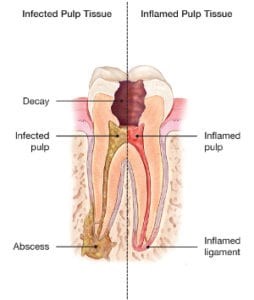
Cause of pulp infection or inflammation
Why microscope is important for root canal treatment?
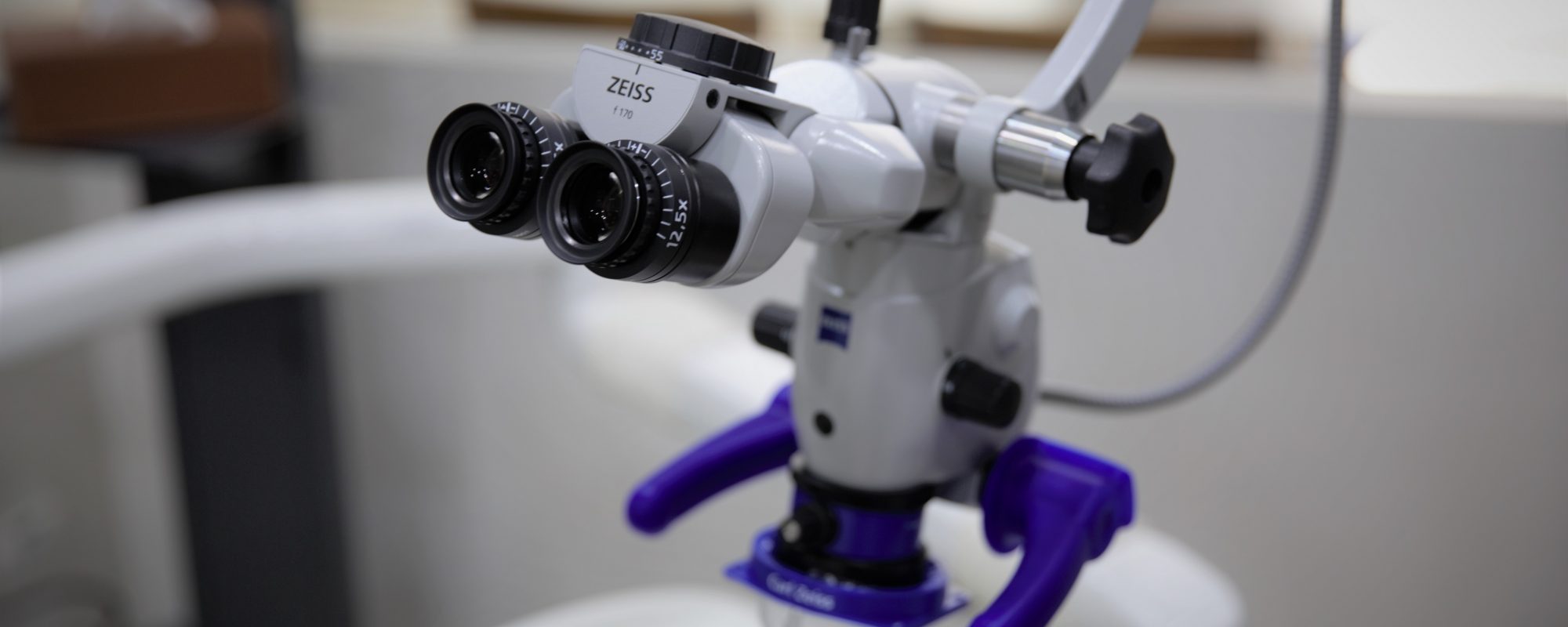
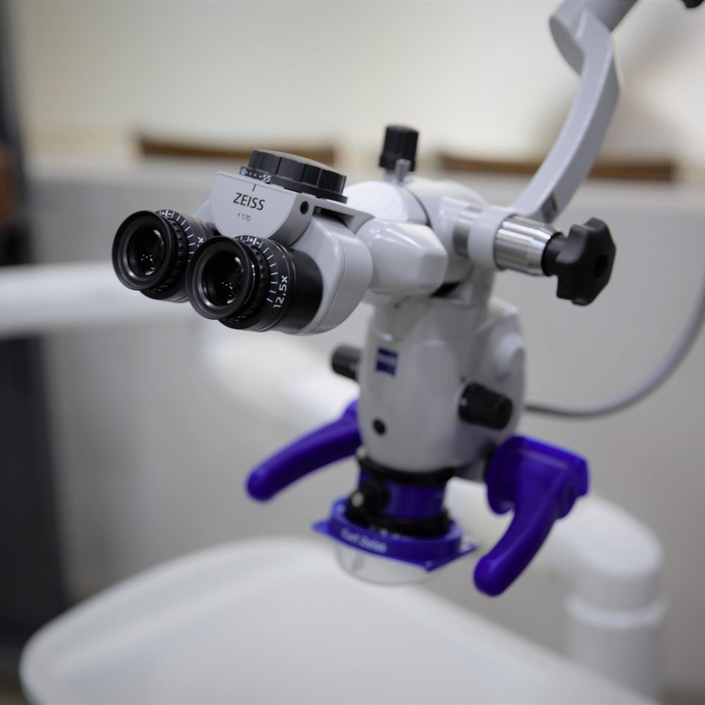
Microscope allows the endodontist to see the inside of a tooth and the root canal system at a magnification of up to 20 times. It enables endodontist to find the tiny canal entry points and branches because teeth often have extra roots or canals that cannot be seen with the naked eye or loupes (glasses with magnification). Sometimes, teeth can have fractures, cracks or other abnormal anatomy. With microscope, these can be detected and treated early. From research, microscope increases success rate of root canal treatment by more than 90 percent.

Procedures for root canal treatment in Bangkok
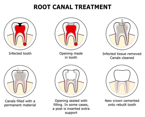
Root canal therapy may involve one or more dental visits. Your endodontist will perform the necessary steps to save your tooth:
- Endodontist will give local anesthesia. Your tooth is numbed for your comfort. A thin sheet of latex or non-latex material called a rubber dam is placed over your tooth to keep it dry. An opening is made through the crown of the tooth into the pulp chamber.
- Your tooth’s nerve or pulp is removed from the tooth. Each root canal is cleaned, shaped and disinfected.
- Your dentist may place medicine in the pulp chamber and root canal(s) to help treat the infection. You may be given antibiotics if the infection has spread beyond the end of the root(s).
- The treated root canals are sealed with a rubber-like material so bacteria can not penetrate to the apex of the root.
- A fiber post may be placed in the root canal to help retain the core material, which supports the crown restoration.
- Finally, your dentist restores the tooth with a crown to strengthen it and improve the way it looks.
Does the root canal treatment hurt?
A local anaesthetic is used and the whole procedure should feel little different to that of having a normal filling although the appointment will usually be longer. There will usually be some pain or discomfort for 3‐4 days following the treatment which can be managed with over the counter analgesics. The tooth may feel slightly tender and “different” for a few weeks following treatment.
What if I don’t have the treatment?
The alternative to root canal treatment is the extraction of the tooth. Once the pulp is damaged, it cannot heal without treatment and it is not recommended to leave an infected tooth in the mouth as it can lead to painful dental abscesses, facial swelling and continued bone loss around the root of the tooth. In some cases, it can lead to death from infection to the blood circulation.
For more information on root canal treatment in Bangkok, to speak with a specialist, or to make an appointment, contact us today.
Microscopic Root Canal Treatment: Precision Endodontic Care to Save Natural Teeth
Root canal treatment is used to save a tooth when the pulp inside the tooth becomes infected, inflamed, or damaged. In some cases, root canal anatomy can be complex, narrow, curved, calcified, or difficult to see with the naked eye. Microscopic root canal treatment uses high magnification and bright illumination to help the dentist treat the inside of the tooth with greater precision.
Microscopic root canal treatment is a modern endodontic approach that allows the dentist or endodontist to see small details inside the tooth more clearly. This can be especially useful for complex cases, retreatment cases, missed canals, calcified canals, cracked teeth, or teeth with persistent symptoms after previous treatment.
The goal is to remove infected or inflamed tissue, clean and shape the root canal system, seal the canals properly, and preserve the natural tooth whenever possible. Although not every root canal case requires a microscope, magnification can improve diagnosis and treatment accuracy in selected situations.
What Is Microscopic Root Canal Treatment?
Microscopic root canal treatment is root canal therapy performed with the help of a dental operating microscope. The microscope provides magnification and focused light, allowing the dentist to see the tooth chamber, canal openings, cracks, blockages, and other fine structures more clearly.
During treatment, the infected or damaged pulp tissue inside the tooth is removed. The root canals are cleaned, shaped, disinfected, and filled with a biocompatible material. After root canal treatment, the tooth usually needs a proper final restoration, such as a filling or crown, to protect it from fracture and restore function.
The microscope does not change the basic goal of root canal treatment, but it helps the dentist work more accurately in small and difficult areas inside the tooth.
Why Use a Microscope for Root Canal Treatment?
Root canals are very small. Some teeth have extra canals, hidden canal openings, curved roots, or calcified spaces that can be difficult to locate without magnification. If infected tissue remains inside a missed canal, symptoms may continue even after treatment.
A microscope can help the dentist identify canal anatomy more clearly and manage complex details that may not be visible during conventional treatment. This is why Microscopic root canal treatment is often recommended for cases that require higher precision.
Magnification may help with:
- Finding hidden or extra root canals
- Locating calcified canal openings
- Managing narrow or curved canals
- Detecting cracks or fractures in the tooth
- Removing old filling material during retreatment
- Identifying separated instruments or blockages
- Improving visibility during complex endodontic procedures
When Is Microscopic Root Canal Treatment Recommended?
Microscopic root canal treatment may be recommended when a tooth has complicated root anatomy or when previous root canal treatment has not resolved the problem. It may also be used when the dentist wants to improve visibility for diagnosis and treatment planning.
Common situations where microscopic endodontic treatment may be useful include:
- Persistent tooth pain after previous root canal treatment
- Root canal retreatment
- Missed canals suspected on X-rays or CBCT scans
- Calcified canals that are difficult to locate
- Curved or narrow root canals
- Teeth with complex anatomy
- Suspected cracks inside the tooth
- Dental abscess related to root canal infection
- Root canal treatment for molars with multiple canals
- Cases where precision is especially important to preserve tooth structure
Not every toothache needs root canal treatment, and not every root canal needs microscopic treatment. A dental examination, X-rays, and sometimes 3D imaging are needed to determine the best approach.
Signs You May Need Root Canal Treatment
A tooth may need root canal treatment when the pulp inside the tooth is infected or inflamed. Symptoms can vary, and some infected teeth may not hurt at all.
You should see a dentist if you notice:
- Persistent toothache
- Pain when biting or chewing
- Sensitivity to hot or cold that lingers
- Swelling around the gum or face
- A pimple-like bump on the gum
- Darkening of a tooth after trauma
- Deep decay or a large cavity
- A cracked or broken tooth
- Previous root canal treatment with recurring symptoms
If infection is present, delaying treatment may allow the problem to spread and may reduce the chance of saving the tooth.
Benefits of Microscopic Root Canal Treatment
1. Better Visibility Inside the Tooth
The main advantage of Microscopic root canal treatment is improved visibility. The microscope helps the dentist see small details inside the tooth that may be difficult to detect with normal vision.
2. Helps Find Hidden Canals
Some teeth, especially molars, may have extra canals. If a canal is missed, bacteria can remain inside the tooth. Magnification can help locate hidden canals and improve the completeness of cleaning.
3. Supports Conservative Tooth Structure Removal
Because the dentist can see the internal tooth anatomy more clearly, microscopic treatment may help preserve healthy tooth structure during access and canal location.
4. Useful for Retreatment Cases
Root canal retreatment can be more difficult than first-time treatment because old filling material, posts, blockages, or missed canals may be present. A microscope can help the dentist manage these challenges more precisely.
5. Helps Detect Cracks and Complications
Some cracks are difficult to see without magnification. The microscope may help identify crack lines or structural problems that affect the tooth’s prognosis.
6. May Improve Treatment Predictability in Complex Cases
While no treatment can guarantee success, Microscopic root canal treatment can provide better visual control in difficult cases, helping the dentist make more informed treatment decisions.
Microscopic Root Canal Treatment vs. Conventional Root Canal Treatment
Both conventional and microscopic root canal treatment have the same purpose: to remove infection from inside the tooth and seal the canals. The difference is the level of visibility and precision during treatment.
Conventional root canal treatment may be effective for many straightforward cases. However, in complex cases, a microscope can help the dentist locate small canals, manage difficult anatomy, and work with greater accuracy.
Microscopic root canal treatment is especially valuable for molars, retreatment cases, calcified canals, suspected cracks, and teeth with persistent symptoms after previous treatment.
The Microscopic Root Canal Treatment Process
1. Consultation and Diagnosis
The dentist will begin by asking about your symptoms, dental history, and previous treatments. The tooth will be examined clinically, and X-rays may be taken. In complex cases, a 3D dental scan may be recommended to evaluate root anatomy, infection, or hidden canals.
2. Local Anesthesia
Root canal treatment is usually performed under local anesthesia. The goal is to keep the tooth and surrounding area numb and comfortable during treatment.
3. Isolation with a Rubber Dam
A rubber dam is commonly used to isolate the tooth. This helps keep the treatment area clean and prevents saliva from entering the root canal system during the procedure.
4. Accessing the Tooth
The dentist creates a small opening in the tooth to reach the pulp chamber and root canals. With the microscope, the dentist can inspect the internal anatomy more clearly.
5. Locating the Canals
The canal openings are located under magnification. This step is especially important when canals are calcified, hidden, or unusually shaped.
6. Cleaning and Shaping
The infected or inflamed tissue is removed. The canals are cleaned, disinfected, and shaped using specialized instruments. The goal is to remove bacteria and prepare the canals for sealing.
7. Filling the Canals
Once the canals are clean and dry, they are filled with a root canal filling material. This seals the canal space and helps prevent bacteria from re-entering.
8. Final Restoration
After treatment, the tooth needs to be restored. Depending on how much tooth structure remains, the dentist may recommend a filling, onlay, or crown. Back teeth often need crowns after root canal treatment because they handle strong chewing forces.
Microscopic Root Canal Retreatment
Root canal retreatment may be needed if a tooth has persistent infection or symptoms after previous root canal treatment. This can happen if canals were missed, the tooth was not sealed properly, a new cavity developed, a crown leaked, or the root canal anatomy was difficult to treat the first time.
During retreatment, the dentist removes old root canal filling material, re-cleans the canals, addresses missed anatomy if possible, and seals the canals again. Microscopic root canal treatment is especially useful in retreatment because visibility is critical for finding hidden problems.
Not every tooth can be retreated successfully. If there is a vertical root fracture, severe bone loss, or poor remaining tooth structure, extraction may be recommended instead.
Microscopic Root Canal Treatment for Calcified Canals
Calcified canals occur when the canal space becomes very narrow or partially blocked by hard tissue. This may happen after aging, trauma, deep fillings, or long-term irritation.
Calcified canals can be difficult to locate and treat. The microscope helps the dentist see subtle color changes, canal traces, and small openings more clearly. In some cases, 3D imaging may also be used to guide treatment.
Microscopic Root Canal Treatment for Molars
Molars often have multiple canals and complex anatomy. Some molars may have extra canals that are easy to miss without magnification. For example, upper molars often have small additional canal anatomy that requires careful inspection.
Microscopic root canal treatment can be especially helpful for molars because it allows the dentist to search for hidden canals and treat the tooth more thoroughly.
Is Microscopic Root Canal Treatment Painful?
Root canal treatment is commonly associated with fear, but the procedure is usually performed under local anesthesia. The purpose of treatment is to remove the source of pain and infection, not to cause pain.
During Microscopic root canal treatment, you may feel pressure or vibration, but you should not feel sharp pain. After the procedure, the tooth may feel tender for a few days, especially when biting. This usually improves as the surrounding tissues heal.
If pain worsens, swelling develops, or symptoms do not improve, you should contact your dentist.
How Long Does Microscopic Root Canal Treatment Take?
The treatment time depends on the tooth type and complexity. A front tooth may take less time than a molar with multiple canals. Retreatment, calcified canals, or complex anatomy may require longer appointments or multiple visits.
Your dentist will explain the expected treatment time after examining the tooth. The priority should be thorough cleaning and proper sealing rather than rushing the procedure.
What Happens After Microscopic Root Canal Treatment?
After treatment, mild tenderness is normal for a short period. You should avoid chewing hard foods on the treated tooth until the final restoration is completed. If a temporary filling is placed, it is important to return for the final restoration as instructed.
Aftercare may include:
- Taking medication as recommended
- Avoiding hard chewing on the treated tooth
- Keeping the area clean with gentle brushing and flossing
- Returning for the final filling or crown
- Attending follow-up visits if recommended
- Contacting the dentist if swelling, severe pain, or bite discomfort occurs
Why the Final Restoration Matters
A successful root canal treatment can fail if the tooth is not properly restored afterward. Once the canals are treated, the tooth needs a strong and well-sealed restoration to prevent leakage, fracture, or reinfection.
For front teeth with minimal damage, a filling may be enough. For back teeth or teeth with large cavities, a crown or onlay may be recommended. The final restoration protects the tooth and helps it function properly in the bite.
Can Microscopic Root Canal Treatment Save Every Tooth?
No treatment can save every tooth. Microscopic root canal treatment can improve visibility and help manage complex cases, but the outcome depends on the tooth’s condition.
A tooth may not be restorable if it has:
- A vertical root fracture
- Severe bone loss
- Extensive decay below the gumline
- Very little remaining tooth structure
- Advanced periodontal disease
- Repeated infection with poor prognosis
If the tooth cannot be saved, the dentist may discuss extraction and replacement options such as a dental implant, bridge, or denture.
Microscopic Root Canal Treatment vs. Tooth Extraction
When possible, saving a natural tooth is often preferred. A treated natural tooth can help maintain chewing function, preserve the bite, and avoid the need for tooth replacement.
However, extraction may be the better option if the tooth has a poor long-term prognosis. The decision should be based on diagnosis, remaining tooth structure, root condition, gum support, cost, treatment time, and patient goals.
Microscopic root canal treatment may be worth considering when the tooth is restorable and the main problem is infection inside the root canal system.
Risks and Limitations
Like any dental treatment, Microscopic root canal treatment has risks and limitations. Possible issues include persistent infection, instrument separation, perforation, missed anatomy, root fracture, post-treatment discomfort, or the need for retreatment or surgery.
Using a microscope can help improve visibility, but it does not eliminate all risks. The dentist will explain the expected prognosis based on your tooth’s specific condition.
How to Maintain a Root Canal Treated Tooth
After treatment, long-term maintenance is important. A root canal treated tooth can last for many years when properly restored and cared for.
- Complete the final restoration as recommended
- Brush and floss daily
- Avoid chewing hard objects such as ice or pens
- Wear a night guard if you grind your teeth
- Attend regular dental checkups
- Treat cavities or gum problems early
- Report pain, swelling, or bite changes promptly
Questions to Ask Your Dentist
Before starting Microscopic root canal treatment, you may want to ask:
- Why do I need root canal treatment?
- Is my case complex enough to need a microscope?
- Do I have hidden or calcified canals?
- Is this first-time treatment or retreatment?
- What is the chance of saving the tooth?
- Do I need a 3D scan?
- Will I need a crown after treatment?
- What happens if symptoms do not improve?
- What are the alternatives to root canal treatment?
Conclusion: Microscopic Root Canal Treatment Helps Treat Complex Teeth with Greater Precision
Microscopic root canal treatment is an advanced approach to endodontic care that uses magnification and illumination to improve visibility inside the tooth. It is especially useful for complex anatomy, missed canals, calcified canals, retreatment cases, molars, and teeth with persistent symptoms after previous treatment.
The goal is to remove infection, clean and seal the root canal system, and preserve the natural tooth whenever possible. While not every tooth can be saved, a careful diagnosis and microscopic technique can improve treatment planning and precision in many challenging cases.
If you have tooth pain, swelling, a previous root canal problem, or a tooth your dentist wants to save, Microscopic root canal treatment may be an important option to consider before extraction.
Frequently Asked Questions About Microscopic Root Canal Treatment
What is Microscopic root canal treatment?
Microscopic root canal treatment is root canal therapy performed with a dental operating microscope. The microscope helps the dentist see small canal openings, cracks, calcification, and complex anatomy more clearly.
Is microscopic root canal treatment better than regular root canal treatment?
It can be especially helpful in complex cases because it improves visibility and precision. However, not every simple root canal case requires a microscope.
When do I need a microscope for root canal treatment?
A microscope may be recommended for root canal retreatment, missed canals, calcified canals, molars, suspected cracks, or teeth with complex root anatomy.
Is the treatment painful?
The procedure is usually performed under local anesthesia. You may feel pressure or vibration, but sharp pain should not be felt during treatment. Mild tenderness afterward is common.
Can microscopic root canal treatment save a failed root canal?
In many suitable cases, microscopic retreatment may help manage a failed root canal by locating missed canals, removing old filling material, and resealing the canals. Success depends on the tooth condition.
Will I need a crown afterward?
Many back teeth need a crown or onlay after root canal treatment to protect them from fracture. The final restoration depends on how much tooth structure remains.
What happens if the tooth cannot be saved?
If the tooth has a poor prognosis, your dentist may recommend extraction and discuss replacement options such as a dental implant, bridge, or denture.