










































Ready to care for your smile with confidence.
For more information or to schedule a consultation with a dentist at Global Dental Complex, please contact us.
📞 Call / WhatsApp: (+66) 065-669-9191
💬 LINE: @globaldentalcpx
🌐 Website: globaldentalcomplex.com/th
Book your consultation today and start planning the treatment that is right for you.

Microscopic Apical Surgery (Apicoectomy) in Bangkok
Microscopic apical surgery, also known as apicoectomy or endodontic microsurgery, is a specialized dental procedure that is used to treat persistent infections or inflammation at the root tip of a tooth, which is also known as the apex. It is a minimally invasive surgical procedure that involves removing a small portion of the apex of the tooth, along with the surrounding infected tissue, to eliminate the source of the problem.
What is Microscopic Apical Surgery?
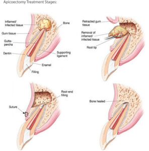
The procedure is typically performed under local anesthesia and involves making a small incision in the gum tissue to access the root of the tooth. A small hole is then drilled into the bone to gain access to the apex of the tooth, and the infected or inflamed tissue is removed using specialized microsurgical instruments. The root tip is then sealed with a filling material to prevent further infection.
When is Apicoectomy Recommended?
Microscopic apical surgery is often recommended when traditional root canal therapy has failed or when there is a persistent infection or inflammation that cannot be resolved through other means. It is also used to treat root fractures, root canals that are blocked or difficult to access, and other complex dental problems.
Benefits of Endodontic Microsurgery Technology
The use of high-powered microscopes and specialized surgical instruments allows for greater precision and accuracy during the procedure, which can lead to better outcomes and faster healing times. It is important to consult with a qualified dental professional to determine if microscopic apical surgery is the right option for your specific dental needs.Specialist Endodontists.
Microscopic Apical Surgery: Saving a Tooth When Root Canal Treatment Is Not Enough
When a tooth has already had root canal treatment but infection or inflammation remains around the root tip, removing the tooth is not always the only option. In many cases, a specialized procedure called Microscopic Apical Surgery may help save the natural tooth by treating the problem directly at the end of the root.
Microscopic Apical Surgery, also known as endodontic microsurgery or apical surgery, is a precise surgical procedure performed near the tip of a tooth root. The dentist or endodontic specialist removes infected tissue, trims the root tip, cleans and seals the end of the root, and allows the surrounding bone and gum tissue to heal.
This treatment is often considered when conventional root canal retreatment is not possible, not suitable, or unlikely to solve the problem. With the help of magnification, microsurgical instruments, and advanced materials, microscopic apical surgery allows the clinician to work in a very small area with greater visibility and control.
What Is Microscopic Apical Surgery?
Microscopic Apical Surgery is a dental procedure used to treat infection or inflammation around the end of a tooth root. The “apical” area refers to the root tip, while “microscopic” refers to the use of a dental operating microscope or high magnification during the procedure.
During the surgery, a small opening is made in the gum near the affected tooth. The infected tissue around the root tip is removed, and a small portion of the root tip may be trimmed. The end of the root is then cleaned and sealed with a biocompatible filling material. This helps block bacteria from re-entering the root canal system from the end of the root.
The goal of Microscopic Apical Surgery is to remove the source of infection while preserving the natural tooth whenever possible.
When Is Microscopic Apical Surgery Needed?
Microscopic Apical Surgery may be recommended when a tooth continues to show signs of infection after root canal treatment. It may also be considered when root canal retreatment is too difficult, risky, or not expected to provide a predictable result.
Common situations where this procedure may be considered include:
- Persistent infection around the root tip after root canal treatment
- A cyst or lesion near the end of the root
- Complex root canal anatomy that cannot be fully cleaned from inside the tooth
- A blocked, calcified, or curved root canal
- A fractured instrument or obstruction inside the canal
- A previous crown or post that makes retreatment difficult
- Root canal retreatment has already failed
- The patient wants to try to save the natural tooth before considering extraction
Not every infected tooth needs surgery. In many cases, non-surgical root canal retreatment may be the first option. Your dentist will evaluate the tooth, X-rays, symptoms, existing restoration, and bone condition before recommending the best approach.
How Is Microscopic Apical Surgery Different from Traditional Apical Surgery?
The main difference is precision. Traditional apical surgery may be performed with direct vision or standard instruments. Microscopic Apical Surgery uses magnification, bright illumination, and microsurgical instruments to improve visibility in a very small surgical field.
This is especially important because the root tip area contains small anatomical details that can be difficult to see with the naked eye. Magnification can help the clinician identify root cracks, small canal spaces, missed anatomy, or other factors that may contribute to persistent infection.
Modern microscopic techniques also allow more conservative removal of tissue and root structure. The aim is to treat the source of infection while preserving as much healthy tooth and surrounding tissue as possible.
Benefits of Microscopic Apical Surgery
1. Helps Preserve the Natural Tooth
One of the biggest benefits of Microscopic Apical Surgery is that it may help save a tooth that might otherwise need to be removed. Keeping a natural tooth can help maintain normal chewing function, support surrounding teeth, and preserve the natural structure of the smile.
2. Directly Treats the Root Tip Area
Some infections remain around the root tip even after root canal treatment. Apical surgery allows the dentist to access the infected area directly, remove diseased tissue, and seal the root end from the outside.
3. Useful When Retreatment Is Difficult
Root canal retreatment may not always be practical. For example, a tooth may have a post, crown, complex canal anatomy, or previous restoration that makes access difficult. In such cases, Microscopic Apical Surgery may provide another path to treat the infection without dismantling the existing restoration.
4. Better Visibility and Precision
The use of a microscope helps improve visibility during the procedure. This allows the clinician to work with greater accuracy in a small area and may help identify details that are difficult to see without magnification.
5. Conservative Surgical Approach
Because microsurgical instruments are small and precise, the procedure can often be performed with a more conservative approach compared with older surgical techniques. This may support better healing and reduce unnecessary tissue trauma.
The Microscopic Apical Surgery Procedure
1. Examination and Diagnosis
The first step is a detailed evaluation. Your dentist will review your dental history, symptoms, previous root canal treatment, and current X-rays. In some cases, 3D imaging may be used to evaluate the root, surrounding bone, and location of infection more accurately.
2. Treatment Planning
Before recommending Microscopic Apical Surgery, the dentist will consider whether the tooth can be saved, whether retreatment is possible, and whether surgery offers a predictable benefit. The condition of the tooth crown, root structure, gum health, and surrounding bone are all important.
3. Local Anesthesia
The procedure is usually performed under local anesthesia. This helps keep the area numb and comfortable during treatment. Your dentist may also discuss other comfort options depending on your needs and the complexity of the case.
4. Accessing the Root Tip
A small incision is made in the gum near the affected tooth. The gum tissue is gently moved aside so the dentist can access the bone and root tip area.
5. Removing Infected Tissue
The infected or inflamed tissue around the root tip is carefully removed. This step helps clean the area and allows the dentist to inspect the root tip under magnification.
6. Root Tip Resection
A small portion of the root tip is removed. This helps eliminate the area where bacteria and complex root canal anatomy may remain. The goal is to remove the problematic portion while preserving as much root length as possible.
7. Root-End Preparation and Filling
The end of the root is prepared with specialized instruments and sealed with a biocompatible root-end filling material. This seal is important because it helps prevent bacteria from leaking out of the root canal system into the surrounding bone.
8. Closing the Gum Tissue
After the root end is sealed, the gum tissue is repositioned and sutured. The area will then begin healing over the following days and weeks.
Is Microscopic Apical Surgery Painful?
Many patients feel anxious about the word “surgery,” but Microscopic Apical Surgery is usually performed with local anesthesia to keep the area numb during the procedure. You should not feel sharp pain while the surgery is being performed.
After the procedure, mild swelling, tenderness, bruising, or discomfort may occur for a few days. These symptoms are usually manageable with aftercare instructions and medication recommended by your dentist. Recovery can vary depending on the tooth location, size of the infection, and individual healing response.
Recovery After Microscopic Apical Surgery
Most patients can return to normal daily activities relatively soon, but it is important to follow post-operative instructions carefully. Proper care helps support healing and reduces the risk of complications.
After Microscopic Apical Surgery, your dentist may advise you to:
- Apply a cold compress during the first day if recommended
- Avoid chewing directly on the surgical area
- Eat soft foods during the early healing period
- Keep the area clean without disturbing the wound
- Avoid smoking, as it can slow healing
- Take medication exactly as prescribed
- Return for follow-up visits and suture removal if needed
You should contact your dental clinic if you experience severe pain, increasing swelling, fever, pus, uncontrolled bleeding, or any symptom that feels unusual.
How Long Does Healing Take?
Soft tissue healing usually begins within days, but bone healing around the root tip takes longer. Your dentist may schedule follow-up X-rays over several months to monitor healing and confirm that the infection is resolving.
It is important to understand that improvement is not always visible immediately on an X-ray. Bone takes time to regenerate, and the healing process depends on the size of the lesion, the quality of the seal, oral hygiene, and the overall condition of the tooth.
Microscopic Apical Surgery vs. Root Canal Retreatment
Root canal retreatment and Microscopic Apical Surgery are both methods used to manage problems after a previous root canal. The best choice depends on the cause of the problem and the condition of the tooth.
Root canal retreatment involves reopening the tooth, removing old filling material, cleaning the canals again, and sealing them from inside the tooth. This may be preferred when the canal system can be accessed safely and the existing restoration can be removed or replaced.
Microscopic apical surgery treats the problem from the root tip side. It may be considered when retreatment is difficult because of a post, crown, obstruction, complex anatomy, or when previous retreatment has not resolved the infection.
Microscopic Apical Surgery vs. Tooth Extraction
Extraction removes the entire tooth, while Microscopic Apical Surgery aims to preserve the natural tooth by treating the infected root tip area. If the tooth is restorable and has enough healthy structure, saving it may be beneficial.
However, surgery is not always the right choice. If the tooth has a vertical root fracture, severe periodontal disease, poor bone support, or insufficient remaining tooth structure, extraction and replacement options such as a dental implant, bridge, or denture may be more appropriate.
Your dentist will help you compare the risks, benefits, cost, treatment time, and long-term outlook of each option.
Who Is a Good Candidate for Microscopic Apical Surgery?
You may be a good candidate for Microscopic Apical Surgery if the affected tooth still has good structural support and the infection is located near the root tip. The tooth should also be restorable, meaning it can support a filling, crown, or other final restoration after treatment.
Good candidates often have:
- A tooth with persistent root tip infection
- A previous root canal treatment that cannot be corrected easily from inside the tooth
- Enough healthy root and bone support
- No severe uncontrolled gum disease around the tooth
- A desire to save the natural tooth when possible
- Overall health that supports normal healing
Your dentist will determine suitability after a full examination. In some cases, another treatment may provide a better long-term prognosis.
Risks and Limitations
Like any surgical procedure, Microscopic Apical Surgery has risks and limitations. Possible risks include swelling, discomfort, infection, delayed healing, sinus involvement for upper back teeth, nerve irritation for certain lower teeth, or failure of the tooth to heal as expected.
The outcome depends on many factors, including the tooth location, root anatomy, quality of previous root canal treatment, presence of cracks, size of the lesion, and the patient’s healing ability. A careful diagnosis is important before deciding whether surgery is the right option.
How to Maintain the Tooth After Surgery
After healing, the treated tooth still needs regular care. Microscopic Apical Surgery addresses the root tip infection, but long-term success also depends on oral hygiene, a proper final restoration, and regular dental checkups.
- Brush and floss daily to control plaque
- Complete any recommended crown or restoration
- Avoid biting very hard objects with the treated tooth
- Attend follow-up appointments to monitor healing
- Visit your dentist regularly for routine checkups
- Tell your dentist if pain, swelling, or biting discomfort returns
Questions to Ask Before Microscopic Apical Surgery
Before starting treatment, it is helpful to ask your dentist or endodontist clear questions, such as:
- Why is apical surgery recommended in my case?
- Is root canal retreatment possible instead?
- What are the chances of saving this tooth?
- Do I have any signs of root fracture?
- Will 3D imaging be needed before surgery?
- How long will healing take?
- What should I expect after the procedure?
- What happens if the tooth does not heal?
Conclusion: A Precise Way to Save a Natural Tooth
Microscopic Apical Surgery is a specialized procedure designed to treat infection at the root tip while preserving the natural tooth. It may be recommended when conventional root canal retreatment is not possible, not suitable, or has already failed.
With the use of magnification, microsurgical instruments, and modern root-end filling materials, this treatment allows the dentist to work precisely in a small area and address the source of infection directly. Although it is not suitable for every case, it can be an important option for patients who want to save a tooth before considering extraction.
If you have a tooth with persistent pain, swelling, or infection after root canal treatment, a detailed evaluation is the best first step. Your dentist can help determine whether Microscopic Apical Surgery, root canal retreatment, or another treatment option is most appropriate for your condition.
Frequently Asked Questions About Microscopic Apical Surgery
Is microscopic apical surgery the same as an apicoectomy?
They are closely related. An apicoectomy refers to removing the tip of the tooth root. Microscopic Apical Surgery is a modern approach that uses magnification and microsurgical techniques to perform the procedure with greater precision.
Why would I need apical surgery after a root canal?
You may need apical surgery if infection or inflammation remains around the root tip after root canal treatment, especially when retreatment is not suitable or cannot fully address the problem.
Can microscopic apical surgery save my tooth?
In many suitable cases, Microscopic Apical Surgery may help save a natural tooth. However, success depends on the tooth structure, root condition, bone support, infection size, and overall oral health.
How long does the procedure take?
The treatment time depends on the tooth location and complexity of the case. Your dentist can provide a more accurate estimate after examination and imaging.
Will I need a crown after microscopic apical surgery?
Some teeth may already have a crown, while others may need a new restoration after treatment. A strong final restoration is important to protect the tooth and support long-term function.
What happens if microscopic apical surgery fails?
If the tooth does not heal or symptoms return, your dentist will reassess the condition. Possible options may include further treatment, monitoring, or extraction and replacement with an implant, bridge, or denture.